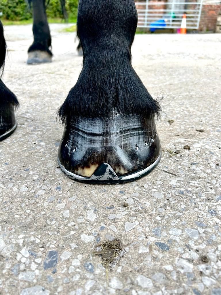
Basic hoof anatomy
In the video opposite, we take you through some basic internal and external hoof anatomy, the video is a basic overview and i have tried to keep it as horse owner friendly as possible, keeping the long anatomical terms to a minimum.
Hope you enjoy.
bones of the lower horses limb (an overview)
In the video opposite, we take you through the bones of the lower limb. Again this video is aimed at educating horse owners so is kept at a basic and easy to understand level.
We hope you enjoy.
The Hoof & Moisture.
by Paul Conroy BSc(hons) AWCF.
The Nourished Hoof Ltd.
The hoof is a multi-dimensional structure that undergoes many different forces at the same time. This means the hoof must be able to resist different forces acting upon it simultaneously. The deeper anatomy of the hoof reveals a complex world of different arrangements and composition of structures that, when combined and in balance, work together to prevent catastrophic failure of the hoof.
The proper functioning of the hoof relies on high-quality hoof care from your skilled farrier, as well as regular maintenance from you, the horse owner. Keep in mind that your farrier checks the hooves every 6 weeks, and by then, it may be too late to address issues that could have been easily prevented with some basic knowledge and daily attention.e
The Hoof:
Keeping things basic, the hoof is made up of 3 basic layers known as stratums, they are known as the Stratum Internum, Stratum Medium and Stratum Externum. Collectively, these 3 layers describe the whole hoof wall, most of the weight borne on the hoof is through the hoof wall.
Stratum Internum: This is the inner most portion of the hoof wall, its primarily the connection between the hard hoof and the sensitive internal structures.
Stratum Medium: This is the main bulk of the hoof wall that contains the three different type of horn that make up the hoof wall.
Stratum Externum: The Stratum Externum is a thin later of waxy and scaly horn that sits on the very outside of the hoof known as The Periople. The Periople has two basic functions.
- It forms a flexible junction between the hard hoof and the soft skin at the coronary band region and prevents bacteria getting in at this vulnerable section of the hoof (A little bit like your cuticles on your nails).
- Its second function is to act as a moisture conveyer to keep the horn that makes up the hoof wall strong and nourished. The Periople is made up of keratinised epithelial cells that absorb moisture in the air and on the hoof wall, they feed the moisture up into the hoof which helps keep the horn cells that make up the hoof wall moist and supple. The horn cells of the hoof wall need to be able to hold up a lot of weight (horses are heavy as anyone who has even been trodden on will know). The hoof bends slightly under load. Obviously, the hoof ability to bend requires the wall to be supple otherwise it will crack.
The Basic principles of hoof moisture.
The hoof can withstand all weather conditions. The hoof likes stable and consistent temperature and moisture levels, what the hoof doesn’t like is changes to these conditions, the problem is that planet earth has seasons, these seasons and different weather conditions would be fine if they were consistent, but we all know they are not. Speaking as a farrier from the UK I can say with certainty that the weather can change not only daily but hourly sometimes. The same can apply to temperature also, the hoof likes slow and gentle temperature changes to give it time to adapt. Drying out or sudden exposure to water can play havoc with the hoof capsule.
Plan Ahead.
Spring/ Summer:
The key to avoiding summer dry cracked or split hooves and shelly feet is to try and maintain the same moisture levels, regardless of the external weather and warmth. As a rough guide, when the external temperature exceeds around 12-15 Degrees Celsius, the hoof begins to lose moisture and dry out. By applying a water-based moisturiser, you can slow down this dry out rate significantly. The warmer it gets, the more it needs moisture to maintain the same moisture level. By flattening the dry out rate and maintaining the same moisture levels the less likely you will experience drying and cracking as well as shoe loss. If you apply a moisture giving cream early and not wait until you notice they are dry and cracking, you will prevent many summer hoof related problems. Only applying a moisture giving cream when the cracks appear is too late.
Our Caffeine enriched daily hoof emollient is specifically designed for spring and summer, its essentially a moisture bomb for the hoof and will significantly slow down the moisture dry out rate in the warm months, the caffeine acts as a stimulant around the coronary band to maximise blood flow to this area where the hoof grows. See the photos on our products page demonstrating this in action.
Autumn/ Winter:
The main problem in the colder seasons is excess moisture and water, a submerged hoof capsule will have no option but absorb water/moisture making it expand quickly and split. Temperature and moisture evaporation isn’t a problem in these seasons, it’s the excess water and muddy fields that pose the threat to hoof health.
Our luxury hoof butter is designed for the winter months, not only does it contain some exquisite oils and organic shea butter, it also contains a water repellent that will resist water ingestion. Our butter is a true cosmetic grade organic shea butter unlike a lot of hoof butters out there that are solidified fats. Because it’s a true butter, it behaves like one, it sticks to the hoof very well and is very resistant to harsh environments, in normal conditions we found it only needs applying every 2-3 days, in harsh conditions, very 1-2 days.
Our hoof products have been tailored for individual seasons, yet they are versatile enough to be effective year-round. While one shines in the summer, the other excels in the winter.
Our Farrier finish daily hoof dressing will work all year round. Not only does it supply an outstanding show shine, it contains no fewer than 5 quality oils all chosen for their strengthening and nourishing properties alongside our eucalyptus essential oil that provides a natural anti-bacterial barrier to the hoof.
For a full description of all our products, please visit our Products page.
To purchase any of our products, please visit our online store.
Hoof Stratums
By Paul Conroy BSc(hons)AWCF
The Nourished Hoof Ltd
The hoof is divided up into 4 stratums of the hoof wall.
The Stratum tectorium.
The stratum tectorium is the very outermost layer of the hoof and is a continuation of the Periople down to the ground bearing border. It is made up of microscopic cells called epithelial cells. These are keratinised cells that appear like dragons scales under the microscope. They help control the moisture intake of the hoof wall and are responsible for the varnish like appearance of the hoof. The act of farriery and the necessity to rasp the hoof wall often removes this layer from the hoof, as does abrasive surfaces.
The Stratum Externum.
The stratum externum refers to the periople. The periople originates from the perioplic corium which sits in the perioplic groove. The periople extends to about 1 inch down the hoof wall before it thins out into the stratum tectorium. The periople is thickest at the heels where it blends in with the frog. The periople serves two functions. Its primary function is to bridge the gap between the skin and the hoof forming a flexible junction that prevents foreign bodies entering the body from that area. Its second function is to work alongside the stratum tectorium to control the moisture content of the hoof wall.
The stratum medium.
The stratum medium refers to the main bulk of the hoof wall. It is the pigmented zone of the hoof containing the horn tubules, inter and intra tubular horn.
The Stratum Medium.
This forms the main bulk of the hoof wall and contains the all 3 types of horn that make up the hoof capsule. it grows from the coronary body in the coronary groove. it begins at the outermost layer of the hoof wall and ends at the un-pigmented zone of the hoof wall.
The Stratum internum.
The stratum internum is the un-pigmented zone of the hoof wall and the insensitive laminae which interdigitate with the sensitive laminae from the laminar corium.
Mud Fever (Cracked Heels)
By
Paul Conroy BSc(hons)AWCF
Mud fever /cracked heels
This condition is something we as farriers see a lot of in the winter months, let’s familiarise ourselves with what it is and what to do.
Mud fever and cracked heels are essentially the same thing. It can also be known as pastern dermatitis or greasy heel, it is softening or cracking of the skin which allows bacteria or other infection causing things in, it typically starts at the heel bulbs and works it’s way up the leg.
Causes
- Excessive exposure to wet conditions that soften the skin allowing infection to seep in
- Skin irritation
- Mites
- Trauma
- Fungus infection
- Congenital disposition such as feathered horses
- Abrasion’s
- Bacterial infection
- Certain soils such as sandy arenas or rough vegetation
- Excessive leg washing
- Bedding that causes irritation
- Incorrect placement or dirty bandaging or over reach boots
- Any disease that lowers the immune system
- Photosensitisation
- Allergies
- Tumours
Signs
Again these can be numerous and I have listed the main ones below
- Lesions on the legs
- Hair loss
- Scaling skin
- Itchy skin
- Crusting over lesions
- Skin folds
- Fluid accumulation
- Serum discharge
- Skin ulcers
- Thick and hardened crusts on skin
- Swelling / pain
- Self trauma such as biting and scratching it’s limbs
- Stamping feet
- Lameness
Diagnosis
If you suspect mud fever then veterinary assistance should always be sought.
A physical examination by the vet should be carried out, they may take hair samples or smears and scans to test for fungal and bacterial presence or mites. Any information about the horses management or living conditions and horse population in the fields that may have mites can help the vet in a diagnosis.
Treatment
The treatment will depend on the cause of the cracked heels / mud fever but the baseline treatment is to remove the infection and allow the skin to heal while treating any underlying condition.
Often hair is clipped away to allow better management of the area, scab removal is generally not recommended but may help in cases of Dermatophilus congolensis as it cannot survive in the presence of oxygen.
Only remove scabs that are soft and ready to fall off.
Rinse well and dry the legs thoroughly using only a clean towel.
An anti bacterial cream may be used daily after washing the legs.
During treatment the horse should be stabled and removed from wet and muddy conditions.
Cleaning the area with a medicated, iodine, chlorhexidine shampoo (always seek veterinary advice beforehand) can promote normal skin bacteria.
A barrier cream to repel water can be used if stabling the horse is not possible, bandages may also prove useful to keep the affected areas clean.
Other treatments may also be prescribed by the veterinary surgeon depending on the specific case which is another reason why veterinary help should always be sought.
Summary
This is another condition that can quickly spiral out of control and can be very painful for your horse, early effective veterinary and owner treatment can have a huge impact in lessening the progression of this condition. Be aware and be proactive.
Cellulitis
By
Paul Conroy BSc(hons) AWCF
Cellulitis
Cellulitis or septic cellulitis is a bacterial infection of the soft connective tissue under your horses skin. It can occur anywhere on the body but commonly occurs in one of the hind legs.
It typically starts with a sudden swelling that is warm and painful to the touch. As the infection progresses, the swelling can spread to affect the whole leg and the horse can develop a fever. Lameness can become so severe that the horse can become non-weight bearing. Fluid may begin draining from cracks in the skin and in some cases the horse may show signs of depression and a loss of appetite. The infection can progress to this point within a few hours.
Some can develop life threatening complications such as laminitis or necrosis (death) of skin tissue.
Causes
The causes are numerous and I have listed them below
- Poor hygiene
- Decrease blood/lymphatic circulation (long term stabled)
- Breaks in the skin
- Following a wound (no matter how tiny the wound)
- Drying and irritation of the skin (Long exposure to mud can cause cracks and breaks in the skin which allow bacteria present on the skin to enter the body)
The above list is by no means exhaustive and there are many other contributing factors.
Signs
- Swelling and pain around the injury (wound) site
- Skin that is hot to the touch
- Sometimes fever in the horse
- Lameness (acute) on the affected limb, sometimes the lameness can occur before the swelling.
Treatment
It is vital you seek veterinary help if you suspect cellulitis as the infection can spiral very quickly and most treatments include antibiotics and anti-inflammatory’s.
The vet will also likely recommend the following
- Keeping the affected area clean and the surrounding hair clipped.
- Possibly cold hosing to reduce the swelling followed by a pressure wrap
- Exercise can be one of the best treatments available (and it’s free!!) this will help the lymphatic system drain the accumulated fluid and improve circulation in the affected area.
Summary
Cellulitis is one of those conditions that can become extremely serious in a very short space of time, with even wetter weather on the horizon and the winter slog about to begin, familiarise yourself with this condition and always seek veterinary assistance if you even suspect it could be cellulitis.
Mallenders & Sallenders.
By
Paul Conroy BSc(hons) AWCF
Mallenders and Sallenders.
Many of you may never have heard of it, unless of course you have the pleasure of owning a traditional type horse with lovely feathers.
Mallenders occurs at the back of the knee and Sallenders occurs at the front of the hock, although they are called different names they are essentially the same thing. It’s easily recognised as scabs or thickened crusty scale in those areas under the feather.
At best it can be uncomfortable for your horse and at worse it can allow a secondary pathogen to cause a bacterial or fungal infection that can and does cause lameness.
It is nearly always seen exclusively in horses with feather. Horses with feather produce more keratin than their non feathered counterparts and it’s this excess of keratin that is responsible for the excess fur and feather, keratin is a protein that is the foundation of skin, hair, hoof etc. this keratin secretes through the skin and if left there to dry will cause these scabs and inflammation.
There is no cure for Mallenders and Sallenders but it’s quite easy to mange if your diligent. Thorough grooming in this area will remove the excess keratin that causes the problem so it’s far less likely to occur, if it does then there are treatments out there (such as equiderma cream) to break down, soften and remove the scabs that cause the problem.
Apply it to the skin and allow it to soften the waxy scale then gently remove it with a wide toothed comb, it can also help to prevent secondary infections.
It’s a condition that we as farriers see and feel a lot in our every day life and can make a horse difficult to shoe because it’s just uncomfortable for them to bend their legs.
I hope this post is useful in better understanding what it is and more importantly, what to do about it.
Foot Abscess's
By
Paul Conroy BSc(hons) AWCF
An Abscess is a local bacterial infection in the hoof.
The infection becomes trapped in the hoof capsule casing pressure to build (much like a tooth abscess).
They are 2 types of abscess depending on where the infection is trapped.
Sub Mural - These are the most painful (in my opinion). The are located behind the hoof wall and are trapped between the hoof wall and the distal phalanx (P3 or Coffin bone).
Sub Solar- These develop under the sole of around the bars.
Lameness can be very fast developing, a horse can go out perfectly sound and be fracture lame within a few hours. If this happens veterinary assistance is required urgently. The abscess needs to be drained. The vet is always the best person to call as they have the necessary equipment to deal with the aftermath of a drained abscess, especially if blood is needed to be drawn to get the abscess.
An abscess is not particularly dangerous or a cause for concern if dealt with promptly and professionally. There isn’t really much you can do to prevent one and it’s often just a case of bad luck if your horse develops one. Certain diseases such as laminitis may make the possibility of an abscess more likely due to the open nature of the feet and stretched white line, this also applies to collapsed or poor quality feet.
Good stable management and hygiene will greatly reduce your risk, picking your feet out and using the brush side to remove the mud and stones will again reduce the risk.
They can vary greatly in depth and position, the deeper more severe abscesses may require some wall drilling as the photo below right shows.
Most abscesses though are relatively superficial and no surgery is required, they are found either sub solar (under the sole) or sub mural (under the wall) from experience, I would say sub mural abscesses cause the most discomfort. The discomfort is due to the pressure build up and once released the horse can show an immediate improvement.
Always listen to the advice of your professional when it comes to aftercare and please don’t cut corners as failure to adhere fully to the instructions can lead to a rapid re-infection and you will need to start the process all over again.
Solar Penetrations
By
Paul Conroy BSc(hons) AWCF
Sub solar penetrations.
I have seen a few of these recently so thought I would do a post explaining the dangers of them and more importantly, what to do in event of discovering your horse has one.
A sub solar penetration is when a foreign body enters the solar or basal area of the foot, the foreign body can be anything from a nail or screw to a sharp stone.
THIS IS A CLINICAL EMERGENCY AND YOUR VET SHOULD BE CALLED IMMEDIATELY.
The first thing to say is if at all possible do not remove the object until you have spoken with the vet. If you do remove the object it is VITAL you make a note of the following.
1. Where it went in.
2. The depth AND angle it went in.
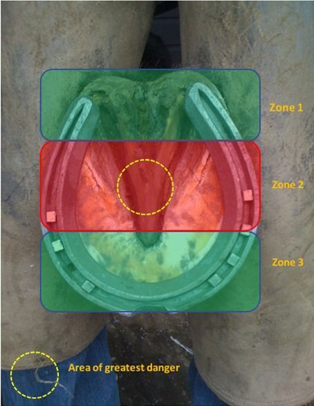
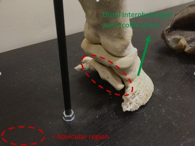
The attached photos (see Below)
I have taken shows the 3 zones of possible penetration, zones 1&3 whilst serious and still needs urgent veterinary assistance are not a serious as zone 2. Sub solar penetrations in zone 2 can be deadly. I have drawn a yellow circle in the centre of zone 2 to show the area of most danger. The anatomy in zone 2 is important to say the least and the biggest danger is the Navicular region, especially the navicular bursa and the Distal Interphalangeal Joint (coffin joint).
If either of these are compromised it can often be fatal for the horse. This is why veterinary assistance is vital. Based on angle and depth the vet can accurately asses whether it can be pulled out and whether x-rays or scans are needed before removal. Upon removal the vet may carry out a street nail procedure, or just dress the wound. They will also make sure your horses' tetanus is up to date. After removal your horse will be on box rest for some time and also your farrier may need to do some surgical/supportive shoeing or a hospital plate may be required, your veterinary surgeon and farrier will discuss this together to formulate a plan based on the individual circumstances of the injury.
First aid.
1. Try to prevent the object going deeper into the foot by placing something (wood ideally) either side of the object, by blocking up the foot in this way the horse can't drive it deeper into the foot if they stand on it.
2. Call the vet immediately, if your not alone, get the other person to call the vet whilst you carry out stage 1.
3. Do not put your horse in a stable with any bedding, the bedding (especially shavings) can and will get between the wooden blocks and drive it deeper into the foot. Stand the horse on level flat ground and wait for the vet.
If you do remove the object in a panic or through vet advice, firstly don't throw the object away, ensure you keep it so the vet can see it, secondly try to stem the bleeding by applying firm pressure and keep the wound clean, sometimes on removal the horse can be immediately better, do not allow the horse to walk around on it or allow it into its bedding as this will make the wound dirty and increase infection risk massively. Be careful removing objects from the frog as due to its high water content and rubbery consistency the hole will close up quickly, ensure you can point to the exact point of entry post removal.
By following these steps and having a deeper understanding of the first aid and the importance of professional help will greatly increase your horses chance of making an uncomplicated recovery from this injury. In most cases your horse will make a full recovery from this injury in time, however, as I stated before, in extreme cases it can be fatal, don't ever leave it to chance. Veterinary medicine and skill/knowledge is higher than its ever been and this injury is treatable in the vast majority of cases.
Understand your role as horse owners so you know exactly what to do to maximise your horse's chance of recovery should you ever have the misfortune to suffer this emergency.


